Article Text

Abstract
Background and aims: The imbalance between effector and regulatory T cells plays a central role in the pathogenesis of inflammatory bowel diseases. In addition to the thymus, CD4+CD25+ regulatory T cells can be induced in the periphery from a population of CD25− T cells by treatment with transforming growth factor β (TGF-β). Here, we analysed the in vivo function of TGF-β induced regulatory T (Ti-Treg) cells in experimental colitis.
Methods: Ti-Treg cells were generated in cell culture in the presence or absence of TGF-β and tested for their regulatory potential in experimental colitis using the CD4+CD62L+ T cell transfer model.
Results: Ti-Treg cells significantly suppressed Th1 mediated colitis on CD4+CD62L+ T cell transfer in vivo, as shown by high resolution endoscopy, histology, immunohistochemistry, and cytokine analysis. Further analysis of in vivo and in vitro expanded Ti-Treg cells showed that exogenous interleukin 2 (IL-2) was crucial for survival and expansion of these cells.
Conclusion: Our data suggest that regulatory Ti-Treg cells expand by TGF-β and exogenous IL-2 derived from effector T cells at the site of inflammation. In addition to Tr1 and thymic CD4+CD25+ T cells, peripheral Ti-Treg cells emerge as a class of regulatory T cells with therapeutic potential in T cell mediated chronic intestinal inflammation.
- TGF-β, transforming growth factor β
- Tregs, regulatory T cells
- Ti-Tregs, TGF-β induced regulatory T cells
- IL, interleukin
- PBS, phosphate buffered saline
- TNF-α, tumour necrosis factor α
- IFN-γ, interferon γ
- RT-PCR, reverse transcription-polymerase chain reaction
- transforming growth factor β
- FoxP3+
- T cells
- experimental colitis
Statistics from Altmetric.com
- TGF-β, transforming growth factor β
- Tregs, regulatory T cells
- Ti-Tregs, TGF-β induced regulatory T cells
- IL, interleukin
- PBS, phosphate buffered saline
- TNF-α, tumour necrosis factor α
- IFN-γ, interferon γ
- RT-PCR, reverse transcription-polymerase chain reaction
The physiological condition of the intestinal mucosa is characterised by maintenance of a constant equilibrium between activation of the mucosal immune system and suppression of mucosal immune responses.1,2 While on the one hand activation of the mucosal immune system is essential to counteract potentially harmful antigens, control of such responses on the other hand is necessary to avoid undesired immune reactions towards self or harmless antigens contained in the intestinal lumen. Recent evidence suggests that loss of this equilibrium is at least partly responsible for the chronic intestinal inflammation observed in patients with inflammatory bowel diseases.1,3,4,5,6,7,8,9,10,11,12
Maintenance of immunological tolerance towards specific antigens in the gut is mainly achieved during maturation of the immune system by specific deletion of autoreactive clones in the process of negative thymic selection.12–14 Furthermore, many lines of evidence support the concept that different subpopulations of regulatory T cells play a pivotal role in maintenance of immunological tolerance in the gut, thereby preventing mucosal inflammation.1,15–17 Among these cells, Tr1 cells and thymus derived CD4+CD25+ regulatory T cells have been shown to suppress colitis in in vivo animal models.18–20 Tr1 cells can be generated in vitro in the presence of interleukin (IL)-10 and their regulatory effect is mediated by secretion of this immunosuppressive cytokine. Moreover, genetically engineered Tr1 cells obtained by retroviral transduction of the IL-10 gene have been recently used for prevention of Th1 mediated colitis in animal models, suggesting the therapeutic potential of cytokine induced regulatory T cells.21,22
Naturally occurring CD4+CD25+ regulatory T cells (Tregs) representing 5–10% of peripheral CD4+ T cells are generated in the thymus in the first days after birth.13 CD4+CD25+ Tregs have been shown to mediate their regulatory function in vitro in a contact dependent manner,23 while in vivo their preventive and therapeutic effect has been linked to expression of IL-10 and transforming growth factor β (TGF-β).17,24 Naturally occurring Tregs constitutively express CD25, GITR, CD103, and CLTA-4 on their surface and, in contrast with Tr1 cells, are characterised by selective expression of the winged helix transcription factor FoxP3.13,25,26 Moreover, experiments using a FoxP3 retroviral vector have demonstrated that expression of this transcription factor is sufficient to direct a genetic programme responsible for acquisition of regulatory properties in CD4+CD25− naïve T cells.13
We27 and others28 have recently demonstrated that CD4+CD25+FoxP3+ T cells can be generated in vitro by polyclonal activation of CD4+CD25−FoxP3− T cells in the presence of TGF-β, thus identifying a novel second class of FoxP3 expressing regulatory T cells. These cells, similar to thymus derived CD4+CD25+ T cells, were shown to act in vitro in a contact dependent manner, thereby suppressing proliferation of naïve T cells in cell culture. In the present work, we have analysed the immunoregulatory capacity of such TGF-β induced CD4+CD25+FoxP3+ T cells (denoted Ti-Treg cells) in vivo using an experimental model of colitis induced by the adoptive transfer of CD4+CD62L+ cells in immunocompromised SCID mice. We found that Ti-Treg cells were able to suppress Th1 mediated chronic colitis induced by adoptive T cell transfer. Moreover, our findings suggest a requirement for exogenous IL-2 provided by an ongoing local immune response for the generation and function of Ti-Treg cells at the site of inflammation where they are required to control pathological immune responses in the colon.
MATERIAL AND METHODS
Animals
Four to six month old BALB/c, SCID, and C57/B6 mice were obtained from the animal facility of the University of Mainz (Germany).
Reagents and media
Antimouse CD3ε (145-2C11) and antimouse CD28 (37.51) antibodies were affinity purified using protein G-Sepharose (Pharmacia Biotech, Freiburg, Germany). Murine recombinant TGF-β1 and murine recombinant IL-2 were purchased from R&D Systems (Wiesbaden, Germany). Serum free medium X-Vivo15 (BioWhittaker, Heidelberg, Germany) or RPMI with 10% fetal calf serum (Perbio Science, Bonn, Germany) containing 2 mM glutamine (BioChrom, Terre Haute, Indiana, USA) was used as indicated. Penicillin/streptomycin was added to all media used (BioChrom).
Endoscopic procedure
For continuous monitoring of colitis, a high resolution mouse video endoscopic system was used, as recently described.29 The endoscopic score of colitis severity (MEICS: range 0–15 points) was based on evaluation of colon translucency (0–3 points), presence of fibrin attached to the bowel wall (0–3 points), granular aspect of the mucosa (0–3 points), morphology of the vascular pattern (0–3 points), and the presence of loose stools (0–3 points).29
Cell isolation
CD4+ T cells were isolated from murine splenocytes using the CD4 cell isolation kit (Miltenyi Biotech, Bergisch Gladbach, Germany). CD4+CD25−/CD4+CD25+ T cells were isolated from CD4+ cells by means of CD25 micro beads kit (Miltenyi Biotech.), while CD4+CD62L+ cells were obtained after positive selection using CD62L beads (Miltenyi Biotech).30
Induction of TGF-β induced CD4+CD25+FoxP3+ regulatory cells (Ti-Treg cells)
Ti-Treg cells were induced as previously described. In brief, CD4+CD25− T cells were plated in anti-CD3 coated wells (10 μg/ml in phosphate buffered saline (PBS) for two hours at 37°C) and cultured in serum free medium in the presence of soluble anti-CD28 (2 μg/ml) and recombinant TGF-β1 (5 ng/ml). In some experiments, recombinant IL-2 (50 U/ml) was added, as specified in the results section. After five days of culture, these cells were magnetically sorted with CD25 microbeads and used in further experiments.
Cytofluorimetry
Day 5 cell cultures were stained for CD25 (BD, Heidelberg, Germany) and FoxP3 (eBioscience, San Diego, California, USA), according to the manufacturer’s instructions. Cells were analysed by means of FASCalibur (BD).
Adoptive transfer and endoscopic screening
To induce colitis in immunocompromised SCID mice, a modification of the originally described protocol was used.30,31 In brief, CD4+CD62L+ cells were isolated from splenocytes of syngenic BALB/c mice, as described above, and 1×10⁶ CD4+CD62L+ cells were transferred in SCID mice alone or in combination with other cells at a ratio of 1:1, as indicated. Mice were screened weekly for signs of colitis by means of a miniaturised colonoscope (Colowiew, Storz, Germany)32 for 6–8 weeks. At indicated time points, colon specimens were collected and analysed. In some experiments, SCID mice were reconstituted with 2×10⁶ Ti-Tregs only generated either from BALB/c or C57/B6 CD4+CD25− cells.
Histological analysis of colon cross sections
Sections were obtained from the removed mice colons and stained with haematoxylin-eosin. The degree of inflammation on microscopic cross sections of the colon was graded by the same pathologist (HAL) in a blinded fashion.
Immunohistochemistry
Immunofluorescence was performed using TSA Cy3 (Perkin Elmer, Heidelberg, Germany) and a fluorescence microscope (Olympus, Melville, New York, USA).33 In brief, cryosections were fixed in ice cold acetone for 10 minutes followed by sequential incubation with methanol, avidin/biotin (Vector Laboratories, Burlingame, California, USA), and protein blocking reagent (Dako, Wiesbaden, Germany) to eliminate unspecific background staining. Slides were then incubated overnight with primary antibodies specific for CD4, CD3, CD11c, IL-6, and tumour necrosis factor α (TNF-α) (all from Santa Cruz, California, USA) or FoxP3 (Alexis Biochemicals, Lausen, Switzerland). Subsequently, the slides were incubated for 30 minutes at room temperature with biotinylated secondary antibodies (Dianova, Darmstadt, Germany). All samples were finally treated with streptavidin-horseradish peroxidase and stained with Tyramide (Cy3 or FITC) according to the manufacturer’s instructions (Perkin Elmer, Heidelberg, Germany). Before examination, nuclei were counterstained with Hoechst3342 (Molecular Probes, Eugene, Ohio, USA).
Reverse transcription-polymerase chain reaction (RT-PCR)
Reverse transcription into complementary DNA was performed using the Moloney murine leukaemia virus reverse transcriptase (Invitrogen, Karlsrhue, Germany). Semiquantitative PCR was performed using the RedTaq PCR reagents (Sigma-Aldrich, Munich, Germany) and the following primers: murine Foxp3, 5′-GGC GAA AGT GGC AGA GAG GTA T-3′ and 5′-AAG ACC CCA GTG GCA GCA GAA-3′; and β-actin, 5′-TGA CGG GGT CAC CCA CAC TGT GCC CAT CTA-3′ and 5′-CTA GAA GCA TTT GCG GTG GAC GAT GGA GGG-3′. PCR products were analysed on 1% agarose gels. Real time PCR was performed using a TaqMan based assay for Foxp3 and HPRT (Applied Biosystems, Applera Deutschland GmbH, Darmstadt, Germany) and ABsolute QPCR Master Mix (ABgene, Hamburg, Germany). FoxP3 expression was calculated relative to the housekeeping gene HPRT on the base of the ddCt algorithm.
Cytokine quantification
IL-10, IL-6, IL-2, and interferon γ (IFN-γ) expression levels in colon tissue were quantified by means of a cytofluorimetry based ELISA system (Flowcytomix, Bender Medsystems GmbH, Austria). In brief, whole proteins were isolated from colon specimens in the absence of detergent. Proteins (100 μg) were immediately used for cytokine determination according to the manufacturer’s instructions. TNF-α levels were analysed by real time PCR using a commercially available TaqMan based assay (Applied Biosystems, Applera Deutschland GmbH, Darmstadt, Germany).
Proliferation assays
Culture medium was RPMI supplemented with 2 mM L-glutamine, 10 IU penicillin, 100 μg/ml streptomycin, and 10% fetal calf serum (heat inactivated at 56°C). CD4+CD25− T cells (5×10⁴) were plated in 96 well round bottom microplates alone or in combination with an equal amount of additional T cells, as indicated in the presence of 1×10⁴ irradiated (4000 rad) antigen presenting cells in a total volume of 0.15 ml of culture medium. In some experiments, 5×10⁴ cells were activated with plate bound anti-CD3 and soluble anti-CD28 in the presence of TGF-β or IL-2. After 72 hours, 0.25 μCi/well of [3H]thymidine were added to the cultures, and after an additional 16 hours thymidine incorporation was assessed by beta scintillation counting.
Statistical analysis
Statistical significance was assessed using the Student’s t test for linear data (weight loss) and the Mann-Whitney U test for non-linear data (endoscopic and histopathological score).
RESULTS
Generation of TGF-β induced regulatory T cells (Ti-Tregs) in vitro
As previous studies suggested that TGF-β stimulation results in the conversion of CD25− T cells into CD25+ T cells,27,28 we tested in an initial series of studies whether such stimulation is sufficient to induce FoxP3 expression and a regulatory capacity under in vitro conditions. Accordingly, we set up an experimental protocol (fig 1A) in which CD25− T cells were activated for five days in the presence or absence of TGF-β. After five days of cell culture, such cells showed induction of FoxP3 mRNA and protein levels by quantitative real time PCR (fig 1B) and immunocytochemistry (fig 1C), respectively. When day 5 cultures were analysed by FACS, 58.2% and 3.1% of cells were CD25+FoxP3+ in the presence and absence of TGF-β, respectively (fig 1D). In order to increase the amount of FoxP3+ cells, day 5 cultures obtained in the presence of TGF-β were magnetically sorted for CD25 and used for further experiments. The so obtained TGF-β induced regulatory T (Ti-Treg) cells showed marked immunosuppressive potential in coculture systems (fig 2A). In fact, newly generated Ti-Treg cells reduced proliferation of freshly isolated CD4+CD25− T cells by more than 80% in a coculture system (p<0.01)(fig 2A). Surprisingly, transfer of Ti-Tregs from Balb/c mice into syngenic immunocompromised C.B-17 SCID mice resulted in markedly reduced FoxP3 expression among CD4+ cells recovered from these mice seven days after transfer (fig 2B, right). This finding suggested that the environment provided by an immunnodeficient host is not sufficient to maintain FoxP3 expression induced in vitro by TGF-β. The same results were obtained when Ti-Tregs were transferred in MHC mismatched conditions (fig 2B, left). These results are in direct contrast with the documented expansion of naturally occurring Tregs when transferred in a lymphopenic host.34 Moreover, T cell receptor stimulation of these cells, as obtained by the MHC mismatch, was not sufficient to expand the population of Ti-Tregs.

(A) Magnetically isolated CD4+CD25− cells were activated for five days with plate bound anti-CD3 and soluble anti-CD28 (2 μg/ml) in presence or absence of transforming growth factor β (TGF-β) (5 ng/ml) in order to generate TGF-β induced regulatory T cells (Ti-Tregs) or preactivated CD4+FoxP3− cells, respectively. (B) Relative expression of FoxP3 mRNA in cells generated from CD4+CD25− T cells activated with anti-CD3 plus anti-CD28 antibodies in the presence or absence of TGF-β. Mean (SD) values from two representative experiments run in duplicate are shown. (C) Immunohistochemistry for Foxp3 on cytospin of CD4+CD25− cells activated with anti-CD3 plus anti-CD28 antibodies in the presence or absence of TGF-β using TSA Cy3 staining system. (D) Day 5 cell cultures were stained with specific anti-CD25 and anti-FoxP3 and analysed by cytofluorimetry. Per cent values are reported in the different quadrants.

Transforming growth factor β (TGF-β) induced regulatory T cells (Ti-Tregs) generated in vitro do not retain FoxP3 expression in a colitis free host. Ti-Tregs generated in vitro from CD4+CD25− T cells via TGF-β (0.5×10⁴) were cocultured with freshly isolated CD4+CD25− T cells (ratio 1:1), and suppression of proliferation induced by plate bound αCD3 and irradiated antigen presenting cells (APCs) was evaluated by means of 3[H]thymidine incorporation relative to that measured in freshly isolated CD4+CD25− T cells cultured alone. CD4+CD25− T cells preactivated in the absence of TGF-β (CD4+FoxP3−) and thymus derived naturally occurring CD4+CD25+ regulatory cells (CD25+ Treg) cocultured with freshly isolated CD4+CD25− cells were used as negative and positive controls, respectively. Thymidine incorporation of Ti-Tregs and CD4+FoxP3− cells alone is also shown. Results represent mean (SD) values from one of three representative experiments. (F) Ti-Treg cells (2×10⁶) generated from CD4+CD25− T cells using BALB/c (right panel) or CB57/B6 (left panel) as donor mice were adoptively transferred into C.B-17/SCID mice (BALB/c syngenic). Four animals in each group were used. Seven days later, CD4+ T cells were isolated and pooled from the spleens of transferred mice. FoxP3 expression was evaluated by semiquantitative reverse transcription-polymerase chain reaction in freshly isolated CD4+ T cells or after 24 hours of activation with plate bound anti-CD3 and anti-CD28 (2 μg/ml) antibodies. FoxP3 expression in unstimulated CD4− cells is shown as a negative control. One of three representative polymerase chain reaction independent experiments is shown.
Ti-Treg cells suppress Th1 mediated colitis induced by adoptive transfer of CD4+CD62L+ T cells in SCID mice
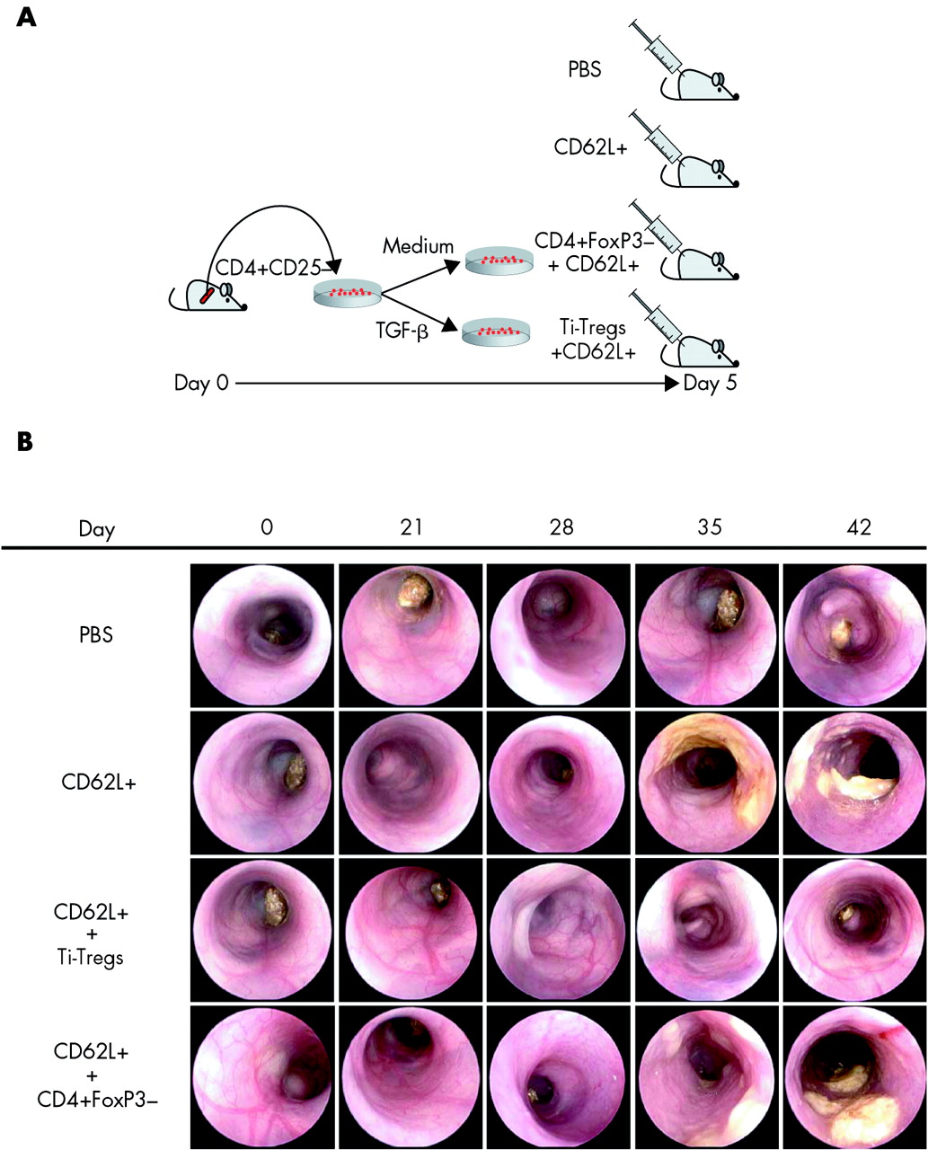
To investigate whether an inflammatory microenvironment is required for Ti-Treg expansion and regulatory capacity maintenance in vivo, we next determined the suppressive effect of these cells in a Th1 mediated colitis model induced by the adoptive transfer of CD4+CD62L+ T cells in SCID mice.30,35 Accordingly, CD4+CD62L+ T cells from BALB/c donor mice were adoptively transferred into SCID mice alone or in combination with CD4+CD25− T cells preactivated in the presence (FoxP3+Ti-Treg cells) or absence (CD4+FoxP3- cells) of TGF-β (fig 3A). Mice were screened weekly by means of a high resolution mini-endoscope,29,32 allowing close monitoring of the development of colitis over time (fig 3B). Within six weeks of cell transfer, mice receiving CD4+CD62L+ T cells alone or in combination with cells precultured in the absence of TGF-β developed severe colitis, characterised by bowel wall thickening, as shown by loss of translucency, bleeding of the mucosa, alterations of the vessel structure, and the presence of massive diarrhoea. In contrast, the colons of mice receiving CD4+CD62L+ T cells together with Ti-Treg cells showed an endoscopic picture undistinguishable from the PBS control group. In fact, endoscopy of these mice showed a translucent bowel wall and a normal vascular pattern. Consistently, a marked reduction in the endoscopic score of colitis severity (MEICS) was noted in mice given CD62L+ wild-type cells plus Ti-Treg cells compared with mice who received CD62L+ CD4+ T cells alone or in combination with CD4+FoxP3− cells (p<0.01) (fig 4).

Transforming growth factor β (TGF-β) induced regulatory T cells (Ti-Tregs) prevent Th1 colitis mediated by the adoptive transfer of CD4+CD62L+ T cells. (A) In order to evaluate the suppressive capacity of in vitro generated Ti-Tregs in an in vivo inflammatory context, 1×10⁶ viable CD4+CD62L+ T cells from BALB/c donor mice were injected intraperitoneally alone or with an equal number of Ti-Treg cells or CD4+CD25− T cells preactivated for five days in the absence of TGF-β (CD4+FoxP3−) in C.B-17 SCID mice. The control group was injected with phosphate buffered saline (PBS) only. Mice were endoscopically screened every seven days over a period of six weeks by means of a miniaturised endoscope. (B) Endoscopic pictures of mice colons from the different groups taken at different time points after the adoptive transfer. Mucosal thickening (as shown by loss of translucency), oedema, and loss of the vascular pattern were seen in mice given CD62L+ T cells or CD62L+ T cells plus CD4+FoxP3− T cells.

Endoscopic score of colitis severity (MEICS) in the different groups was based on evaluation of colon thickening, mucosal granularity, and alteration of the vascular architecture, and on the presence of fibrin or diarrhoea. The scores obtained in the different groups were analysed using the Mann-Whitney U test and a significant difference (p<0.01) was observed between the group of animals reconstituted with transforming growth factor β induced regulatory T cells (Ti-Tregs) and those receiving either CD62L+ cells alone or together with CD4+FoxP3− cells. The horizontal bars represent the median of the endoscopic scores obtained from 9–10 animals per group. Data were pooled from two independent experiments. PBS, phosphate buffered saline.
Consistent with the endoscopic data, reconstituted mice displayed a significant (p<0.01) weight reduction in the groups receiving CD4+CD62L+ T cells alone or in combination with cells preactivated in the absence of TGF-β (18% and 24% of the original weight, respectively) six weeks after transfer while mice receiving CD4+CD62L+ T cells plus Ti-Treg cells or PBS treated mice showed no weight loss (fig 5A). Furthermore, macroscopic analysis of the colons at the end of the experiment showed that cotransfer of Ti-Treg cells suppressed colonic inflammation whereas shortening of the colon was observed in mice cotransferred with CD4+FoxP3− control T cells (fig 5B). Finally, histological analysis of specimens collected from these mice confirmed the endoscopic findings (fig 5C, D). In fact, histological colitis activity analysed in the different groups showed a significant difference (p<0.01) between the groups of animals receiving Ti-Tregs in comparison with animals reconstituted with either CD62L+ cells alone or in combination with CD4+FoxP3− cells (fig 5D). Sections from mice reconstituted with CD4+CD62L+ T cells plus Ti-Treg cells showed no hypertrophy of the colon mucosa and a conserved crypt structure with maintenance of goblet cells in the presence of only a few cells infiltrating the lamina propria. In contrast, mice reconstituted with CD4+CD62L+ T cells plus control cells showed mucosal hypertrophy, high cell infiltration of the lamina propria, and distortion of the crypts (fig 5C). These data are consistent with an in vivo suppressive effect of TGF-β induced regulatory T cells on the development of colitis induced by transfer of naïve cells into immunocompromised mice.

(A) After six weeks from the initial transfer, mice receiving CD62L+ cells alone or in combination with cells preactivated in the absence of transforming growth factor β (TGF-β) (CD4+FoxP3−) showed more than 25% reduction of their original weight while no significant weight loss was observed in animals treated with TGF-β derived regulatory T cells (Ti-Tregs). Data were pooled from two independent experiments. The horizontal bars indicate the average weight loss or gain in individual groups (*p<0.01, Student’s t test). PBS, phosphate buffered saline. (B) Representative macroscopic pictures of the colons isolated from the four different groups. (C) Two representative sets (m1, m2) of haematoxylin-eosin stained colonic sections obtained from the indicated groups. Severe inflammation was noted in mice reconstituted with CD62L+ T cells or CD62L+ T cells plus CD4+FoxP3− T cells. (D) Histopathological scores obtained in the different groups were analysed and evaluated for potential differences using the Mann-Whitney U test (*p<0.01). Data were pooled from two independent experiments.
In vivo expansion of Ti-Treg cells in the colon of reconstituted mice
Cotransfer of Ti-Treg cells resulted in significant upregulation of FoxP3 expression in the colon mucosa in comparison with animals reconstituted with either CD62L+ cells alone or in combination with CD4+FoxP3− cells (fig 6A). Moreover, as shown in fig 6B, we observed an inverse correlation (r = −0.89) between FoxP3 expression at the colon level and histological colitis activity (p<0.001), suggesting that local expansion of Ti-Treg cells occurs on cell transfer in mice resulting in protection from colitis. Consistently, a large number of CD4+ FoxP3 expressing cells could be identified in the colon of mice given Ti-Treg cells compared with mice cotransferred with CD4+ FoxP3− T cells (fig 6C). The few FoxP3+ cells detected in the colons of mice reconstituted with CD62L+ and FoxP3− cells can be attributed to a subpopulation of FoxP3+ CD62L+ cells contaminating the original preparation or alternatively to peripheral induction of regulatory cells not sufficient to control colitis development, as previously reported.36 Taken together, these data suggest sustained suppression of Th1 mediated inflammation over six weeks by colonic Ti-Treg cells expressing FoxP3.

Histopathological score correlates with accumulation of FoxP3+ cells. (A) Relative FoxP3 expression was evaluated in the mouse colons of the different groups (n = 5). Significant upregulation of FoxP3 (**p<0.01) was observed between the group reconstituted with transforming growth factor β derived regulatory T cells (Ti-Tregs) compared with those receiving either CD62L+ cells alone or in combination with CD4+FoxP3−. Bars indicate mean (SD) relative FoxP3 expression in the indicated groups. PBS, phosphate buffered saline. (B) FoxP3 expression in animals from the Ti-Tregs group (triangles) and the CD4+FoxP3− group (circles) was analysed for correlation with the corresponding histological colitis activity score. The Spearman correlation test showed an inverse correlation (r = −0.89), with p<0.001. (C) Representative double staining for FoxP3 (red) and CD4 (green) on colon cryosections obtained from the different groups, as indicated. Four colons per group were analysed.
TGF-β induced Ti-Tregs prevent accumulation of CD4+ and CD11c+ cells in the lamina propria
To obtain further mechanistic insights into Ti-Treg mediated suppression of intestinal inflammation, cryosections obtained from the different groups were stained with anti-CD3 and anti-CD4 antibodies. These stainings showed significant CD3+ and CD4+ T cell infiltration in mice reconstituted with CD4+CD62L+ T cells alone or in combination with cells preactivated in the absence of TGF-β, while cotransfer of Ti-Treg cells prevented such accumulation of CD4+ T cells in the mucosa (fig 7). Furthermore, the group reconstituted with Ti-Treg cells showed a reduction in dendritic cell infiltration compared with the control group, as indicated by staining with anti-CD11c. In addition, there was a marked reduction in staining for proinflammatory cytokines produced by dendritic cells and T cells such as IL-6 and TNF-α (fig 5), implying that Ti-Treg cells exert their regulatory function in vivo by preventing expansion and activation of both antigen presenting cells and CD4+ T cells.

Transforming growth factor β derived regulatory T cells (Ti-Tregs) prevent accumulation of inflammatory cells. Immunohistochemistry for CD3, CD4, interleukin 6 (IL-6), tumour necrosis factor α (TNF-α), and CD11c on cryosections obtained from colon specimens of the different groups, as indicated.
In addition, analysis of cytokine expression by cytofluorimetry based ELISA assay and real time PCR showed a significant reduction in colonic IFN-γ, IL-6, and IL-2 proteins and TNF-α transcripts in mice treated with Ti-Tregs that was accompanied by an increase in IL-10 expression (p<0.01) compared with the control groups (fig 8A). Taken together, these data suggest that TGF-β induced Treg cells suppress colitis activity in vivo by reducing both CD4+ and CD11c+ cell infiltration in the colonic lamina propria and that this effect is accompanied by downregulation of the proinflammatory cytokines IL-2, IL-6, TNF-α, and IFN-γ as well as IL-10 secretion. Moreover, FoxP3 positive cells were clearly identified at the end of the experiment after six weeks (fig 6C), suggesting that Ti-Treg cells may expand and maintain their regulatory potential during an ongoing inflammatory process in vivo. Consistent with this concept, IL-2, expressed at significantly high levels in the colon mucosa of inflamed mice (fig 8A), was able to overcome in vitro TGF-β induced Ti-Treg anergy, as indicated by 3H thymidine incorporation (fig 8B), and such proliferation was accompanied by an increase in relative FoxP3 expression (fig 8C). Therefore, we can conclude that inflammation derived IL-2 is at least in part responsible for in vivo expansion of Ti-Tregs.

Inflammation derived interleukin (IL)-2 sustains transforming growth factor β derived regulatory T cell (Ti-Tregs) expansion and colitis suppression, determining an autoregulatory loop. (A) Accumulation of inflammatory cells prevented by cotransfer of transforming growth factor β (TGF-β) induced Ti-Tregs was accompanied by downregulation of IL-2, IL-6, interferon γ (IFN-γ) protein, and tumour necrosis factor α (TNF-α) messenger as well as upregulation of IL-10 protein, as shown by cytofluorimetric based ELISA assay and real time polymerase chain reaction, as indicated, performed on colonic specimens obtained from the groups treated with phosphate buffered saline (PBS), CD62L+, CD62L+/Ti-Tregs, and CD62L+/CD4+FoxP3−. **p<0.01 between the CD62L+/Ti-Tregs group and both the CD62L+ and CD62L+/CD4FoxP3− groups (Student’s t test. (B) 3H thymidine incorporation shown by Ti-Tregs (CD4+CD25− T cells activated in the presence of TGF-β) and CD4+CD25− T cells, activated in the presence or absence of exogenous IL-2 (50 U/ml). Results represent mean (SD) values of two independent experiments run in triplicate. (C) Relative FoxP3 mRNA expression in Ti-Tregs generated from CD4+CD25− in the presence or absence of IL-2 (50 U/ml). Mean (SD) values from two experiments run in duplicate are shown.
DISCUSSION
In the present study, we have analysed the functional capacity of TGF-β induced CD4+CD25+FoxP3+ T (Ti-Treg) cells and determined the molecular signals required for their generation and functional stability. We observed that Ti-Treg cells suppressed Th1 colitis induced by the adoptive transfer of CD4+CD62L+ T cells thus defining a class of peripherally induced FoxP3+ T cells for suppression of chronic intestinal inflammation.
The protective effect of Ti-Treg cells on colitis activity was mirrored by the reduction in histopathology scores and accompanied by increased FoxP3 expression in lamina propria infiltrating cells. This evidence suggests that the FoxP3+ fraction of the Ti-Tregs culture generated in vitro migrates in the periphery, suppressing activation of colitogenic cells. Nevertheless, the investigative tools available to date do not allow us to completely exclude the contribution of the small fraction of FoxP3− cells present in the same cultures to the suppressive effect obtained in vivo by transfer of Ti-Tregs cells. Interestingly, Ti-Treg cells were not only able to prevent CD4+ T cell accumulation in the lamina propria, but also accumulation of CD11c+ dendritic cells. At the cytokine level, expression of IL-2, IL-6, TNF-α, and IFN-γ was suppressed, while expression of IL-10 was induced in the lamina propria. These data on peripherally induced Ti-Treg cells are consistent with those by Powrie and colleagues on naturally occurring (thymus derived) CD4+CD25+ regulatory cells in experimental colitis.15–17 Thus these findings suggest that thymus derived Treg cells as well as peripherally induced Ti-Treg cells have potent anti-inflammatory functions in chronic colitis in vivo. Consistently, both thymus derived and peripherally induced regulatory T cells (Ti-Treg) exert their effect not only by suppressing accumulation of T cells but also by modulating innate immunity via dendritic cells.37 Furthermore, downregulation of IL-2 observed in animals treated with Ti-Treg cells suggested diminished IL-2 secretion by target cells in a way similar to IL-2 suppression exerted by thymus derived CD4+CD25+ regulatory cells on target cells. Thus our in vivo data support a model in which Ti-Treg cells represent a peripherally induced equivalent to thymus derived CD4+CD25+ Treg cells.
Ti-Treg cells appear to require bystander cells during inflammation to survive and expand in vivo, as transfer of these cells alone in alymphocytic hosts resulted in a complete loss of FoxP3+ T cells within one week. This is in contrast with the observation that naturally occurring Tregs undergo homeostatic expansion in alymphocytic hosts.34 Restimulation in vitro of these cells after reisolation from their SCID hosts failed to reinduce suppressive activity and FoxP3 expression. The TCR engagement, as obtained in MHC mismatch conditions, but in absence of graft versus host disease,38 was not sufficient to sustain expansion of the Ti-Tregs transferred, as indicated by loss of FoxP3 expression observed in SCID mice reconstituted with CD57/B6 Ti-Tregs alone. This finding is further supported by stimulation experiments in which Ti-Treg cells starved for 24 hours showed downregulated FoxP3 expression and lost regulatory activity that could not be rescued by restimulation (unpublished data). Thus our data provide strong evidence for a reversible phenotype of Ti-Treg cells in vivo as well as in vitro, in contrast with the stable phenotype of naturally occurring CD4+CD25+ Treg cells.
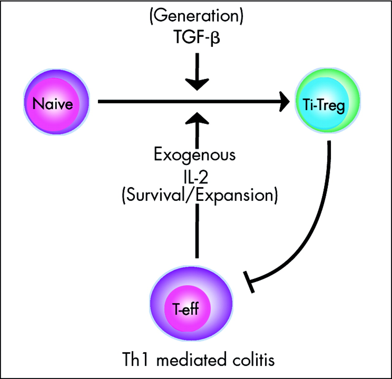
In addition to the well known thymus derived CD4+CD25+ T cells and Tr1 cells, the present study identified Ti-Treg cells as a class of inducible regulatory T cells able to suppress experimental colitis. On the basis of our findings, we propose a model in which naturally occurring CD4+CD25+ regulatory T cells generated in the thymus are responsible for the stable maintenance of tolerance towards T cell clones that are restricted to self antigens or harmless antigens, which have escaped the negative selection process. In contrast, regulatory T cells induced in the periphery (Ti-Treg) would represent a highly adaptive population of regulatory cells sharing with the thymus derived CD4+CD25+ Treg cells expression of FoxP3 and the suppressive capacity but strictly dependent on the presence of exogenous factors such as TGF-β and IL-2 for their development and survival (fig 9). The Ti-Treg population would therefore enable the immune system to turn off inflammatory responses once the harmful antigens are eliminated, thereby avoiding perpetuation of an undesired exaggerated inflammatory response. Moreover, suppression of the ongoing inflammatory process would determine contraction of the pool of the newly generated regulatory cell, so releasing the mucosal immune system by their suppression and making it ready for a new antigenic challenge.
Role of inflammation derived interleukin 2 (IL-2) on expansion of transforming growth factor β derived (TGF-β) regulatory T cells (Ti-Tregs) and the autoinhibitory loop determined by Ti-Tregs mediated IL-2 inhibition on effector cells (T-eff).
Identification of the specific cytokine signals required for generation and functional stability of Ti-Treg cells opens new avenues for therapy of chronic intestinal inflammation. In particular, ex vivo generated TGF-β induced Tregs emerge as a novel approach for the therapy of inflammatory bowel diseases.
REFERENCES
Footnotes
-
Published online first 14 September 2005
-
The work of MFN was supported by grants from the Deutsche Forschungsgemeinschaft and the SFB432
-
Conflict of interest: None declared.